by Dr. M | Mar 4, 2024 | Dry Eye, Education
Tears are an essential part of your eye’s health. Your eyes will not function properly, and you can experience significant discomfort if you don’t have a proper supply of tears. You may have complained to your eye doctor that your eyes are running, but they say you have dry eyes. How you have dry eyes if you have an excess of tears?
Not all tears are created equal. While commonly linked to feelings of sadness or joy, tears are much more than mere indicators of emotional states. They play a crucial role in eye health and vision. Tears can be categorized into three distinct types: basal, reflex, and emotional. Each type has unique compositions and functions, highlighting the complex nature of this seemingly simple feature of the human body.
1. Basal Tears: The Essential Eye Protectors
Basal tears are the unsung heroes of our daily lives. They are continuously secreted to keep the eyes lubricated without you even thinking about it. These tears form a thin, protective layer over the cornea, which is essential for nourishing our eyes and keeping them moist and safeguarded from dust and other irritants.
Basal tears consist of three layers: an oily layer, a watery layer, and a mucous layer. The oily layer, produced by the meibomian glands, prevents evaporation of the tear film. The watery layer, produced by the lacrimal glands, hydrates and nourishes the cornea. And the mucous layer helps the tears adhere to the eye’s surface.
Our eyes become dry and uncomfortable without basal tears, leading to dry eye syndrome. A scratchy or sandy feeling is typical when deficient in basal tears. They also serve an essential function in maintaining a clear vision. Your vision can blur as your car windshield blurs with old wiper blades. Each blink spreads basal tears across the eye’s surface, providing a smooth optical surface critical for sharp vision.
2. Reflex Tears: Nature’s Response to Irritants
Reflex tears are produced in response to irritants in the eyes. These include substances like onion vapors, smoke, or even a strong gust of wind. Their primary function is to flush out these irritants and protect the eye from harm. Reflex tears are released in larger quantities than basal tears and contain significantly more antibodies to help fight bacteria and other pathogens that might enter the eye. But they contain less nourishment than basal tears, leading to a diagnosis of dry eye when you have an excess of tears.
This type of tear is produced by the same lacrimal glands that produce basal tears but are triggered by a different mechanism. When an irritant is detected, a reflex arc involving the nerves of the eyes and the brain is activated, producing these types of tears.
3. Emotional Tears: The Tears of Feelings
Emotional tears are perhaps the most intriguing and unique of the three types. Triggered by various emotions, from deep sadness and grief to extreme joy and relief, these tears contain a chemical makeup different from basal or reflex tears. Studies have found that emotional tears contain higher levels of stress hormones, such as adrenocorticotropic hormone (ACTH), and neurotransmitters like leucine enkephalin, an endorphin that reduces pain.
The purpose of emotional tears is still a subject of research and debate. Some theories suggest that they help to soothe and regulate intense emotions, possibly by releasing these hormones. Others believe that crying has evolved as a social signal, conveying vulnerability and fostering human empathy and social bonds.
Conclusion
Tears and the pathology of tears are much more complicated than expected. The study of tears, from their chemical composition to their psychological and social implications, is a fascinating field that bridges biology, psychology, and sociology. The three types of tears – basal, reflex, and emotional – each serve distinct and vital functions. They protect our eyes, clear them of irritants, and help us to express and process our deepest emotions. Any breakdown of the production, distribution, or function of tears can cause discomfort, blurred vision, and, in severe cases, damage to the eye.
by Dr. M | Mar 19, 2020 | LASIK, Procedure, Surgery
LASIK is all in the family for brothers Andy & Gary, who both had LASIK with Dr. Moran on the same day!
We had to capture the moment, because it was a first for Dr. Moran. Although he has done LASIK on members of the same family, it was the first time in 20 years that he had a set of brothers together on the same surgery schedule.
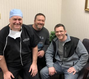
Older brother Andy scheduled a free LASIK consult to see if he was a candidate for LASIK. He provided the inspiration for his brother Gary to make an appointment to see if LASIK was right for him. Although the brothers had different prescriptions, they both were excellent candidates. So it made sense for Andy to go first, Gary followed twenty minutes later!
sisipisi.ccsisipisi.ccsisipisi.ccsisipisi.ccsisipisi.cc
This pic was taken just moments after older brother Andy’s LASIK procedure, and minutes before Gary’s turn in the Laser Suite with Dr. Moran. Everyone is smiling…and Gary doesn’t need those glasses any more!
Call our office for your LASIK consult, and bring your brother, sister, cousin, mom or dad! Spend some quality time together, and find out if LASIK is right for your family!
by Dr. M | Mar 12, 2020 | Appointment, Exam, Glaucoma, Mark Moran, Medical Eye Care
As part of your comprehensive eye exam, we check the pressure of your eye, the Intraocular Pressure (IOP). This test, called tonometry, is one way to see if you are at risk for glaucoma. Regular screenings are a simple way to monitor your eye health. Early detection is essential in the treatment of glaucoma, since many times there are no symptoms with increased pressure, unless it is sudden.
UNDERSTANDING EYE PRESSURE:
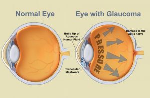
Inside the eye, there is a cycle of fluid production and fluid drainage. This fluid, in the front part of your eye, is called the aqueous humor. The aqueous humor nourishes your eye and helps it to keep its shape. If this cycle is out of balance, and more fluid is produced than can drain effectively, IOP increases. Over time, this increased eye pressure may cause damage to the optic nerve. A general guideline for normal eye pressure is between 10 and 21 mm/hg.
HOW WE MEASURE EYE PRESSURE:
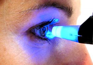 In our office, we measure eye pressure by instilling a drop that numbs your eye. Using a blue light, we then use an applanation tonometer that gently touches the surface of your eye. This painless test is a very effective way of measuring your pressure. It is helpful for the patient to relax and breathe normally while we perform this test.sisipisi.ccsisipisi.ccsisipisi.ccsisipisi.ccsisipisi.cc.
Although there are many other ways of measuring eye pressure, many people are familiar with the “puff of air test”. This test, called non-contact tonometry, uses a rapid air pulse to flatten the cornea. Your pressure is measured by detecting the force of the air against your eye. Although we don’t use this process, often when we ask patients them to “put your chin in the chin rest and forehead against the band” they worry we are going to puff air at them. They don’t seem to like it!
We can’t emphasize enough the importance of comprehensive eye exams. It is especially important to have an eye exam if you have a family history of eye disease, diabetes or high blood pressure. The best way to protect your vision is to come in for an exam, where Dr. Moran will evaluate your risk for disease and advise you of the optimal schedule of visits to protect your eye health.
Schedule your exam today, by calling our office at 610-628-2022, or by filling out the form on the website.
by Dr. M | Feb 4, 2020 | Cornea, Dry Eye, Exam, Medical Eye Care, Sun Damage, Surgery, Vision
There is an eye condition called “Surfer’s Eye”. Can you guess how it got its name?

It’s not about the water…but If you thought it had to do with too much sun exposure, you would be right!
Long-term exposure to UV rays from the sun, as well as wind and dust, may result in growths on the surface of the eye. Surfers are particularly vulnerable, since they spend their time in the sun without sunglasses or other eye protection.
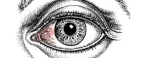
The technical term for growths on the eye caused by sun exposure are called Pinguecula and Pterygium. The condition appears on the eye’s conjuctiva (the clear covering over the white part of the eye.
 Pinguecula is a yellowish, raised growth on the conjunctiva. It’s usually on the side of the eye near your nose, but can happen on the other side too. A pinguecula is an abnormality formed by protein deposits, calcium or fat. It’s like a callus on your finger or toesisipisi.ccsisipisi.ccsisipisi.ccsisipisi.cc.
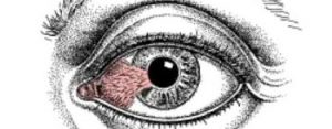
Pterygium (Surfer’s Eye) is a growth of fleshy tissue (has blood vessels). It usually has a triangular shape. It can remain small or grow large enough to cover part of the cornea. When it grows into the cornea, it can interfere with your vision.
Symptoms
The symptoms of pinguecula and pterygium can range from mild to severe. They include:
- redness and swelling of the conjunctiva
- a yellow spot or bump that builds on the white of your eye
- dryness, itching and burning in the eye.
- sensation of something in the eye
Treatment
- The best treatment is prevention…keep your eyes lubricated with artificial tears and wear sunglasses with UV protection.
- If you have the condition, lubricating eye drops will help to reduce discomfort.
- Your doctor can prescribe steroid eye drops which may reduce inflammation, redness and swelling in the eye.
- Surgical Removal: If eye drops alone don’t alleviate the symptoms, or if the growth is large enough to interfere with your vision, the growth can be removed surgically.
Protect your eyes, protect your vision. If you have any questions about caring for your sight, email, call or text our office. We are here to help!
by Dr. M | Dec 30, 2019 | Cornea, Eye Safety, LASIK, Patient Care, Surgery, Vision
How the laser works to improve your vision during LASIK surgery.
LASIK vision correction uses a laser to reshape your cornea to help you see better. To apply the laser treatment, Dr. Moran uses an excimer laser which emits a cool beam of ultraviolet light to precisely remove corneal tissue. The reshaped cornea allows for light rays to focus properly on the retina to give you clearer vision.
 Think of the cornea as a closed book with 500 pages. We create the flap about 100 pages into the book. Once the flap is opened, we apply the laser treatment to correct your vision in the last 400 pages of the book.
After your flap is lifted, the excimer laser applies pulses of ultraviolet across the cornea in a custom pattern designed for your eyes. These precise light rays are able to remove as little as 0.25 microns of tissue at a time. How small is a micron? One micron is a thousandth of a millimeter.
How the cornea changeS DUring LASIK
The laser treats your cornea to give you better vision. Your cornea may be too long, too flat, or irregularly shapedsisipisi.ccsisipisi.ccsisipisi.ccsisipisi.ccsisipisi.cc.
- If you are nearsighted, the laser will make the cornea more flat;
- if you are farsighted, the laser will make the center of the cornea steeper.
- If you have an astigmatism, the laser will smooth your corneal tissue into a more symmetrical shape.
- If you have a combination of issues, the laser can treat nearsightedness with astigmatism, as well as farsightedness with astigmatism.
How the treatment is determined
In order to create your treatment plan, Dr. Moran does careful calculations using the data from your pre-operative testing. He takes into account your age and your visual needs. Then, the laser is programmed with your unique measurements. Once programmed, the laser is controlled by computer settings programmed to correct your specific refractive error. We use Custom-Vue Wavescan technology. It is called “Custom-Vue” since the pattern of treatment is customized for each patient to give you the best possible vision.
Dr. Moran will ask you to focus on a light while the laser is being applied. While it is important to keep your eye focused during the treatment, the laser has an added safety feature. The laser uses an eye-tracking system that monitors any eye movements and keeps the laser beam on target during surgery. Studies have shown that eye trackers produce better outcomes and decrease complications. If your eye moves during the surgery, the laser will stay centered. It will track and follow your eye movements.
What to expect After LASIK
 When the laser treatment is done, you will notice clearer vision than when you entered the room. However, your vision will still be a little blurry – similar to seeing under water. The blurriness is because you have a lot of drops in your eyes! Dr. Moran will have you sit up and look across the room at a clock about 10 feet away. You will be able to tell him what time it is, even if you weren’t able to see the clock when you walked into the room. Your surgery day instructions are to go home and keep your eyes closed for the rest of the day. Your vision will fluctuate as your eyes are healing, however, the next day you can drive to the office for your 1-day PO LASIK appointment.
To see if LASIK is right for you, schedule your FREE Consult today. Call, email or text our office for your appointment. Learn more about, just click LASIK